Neurolymphomatosis detected by 18F-FDG PET/CT
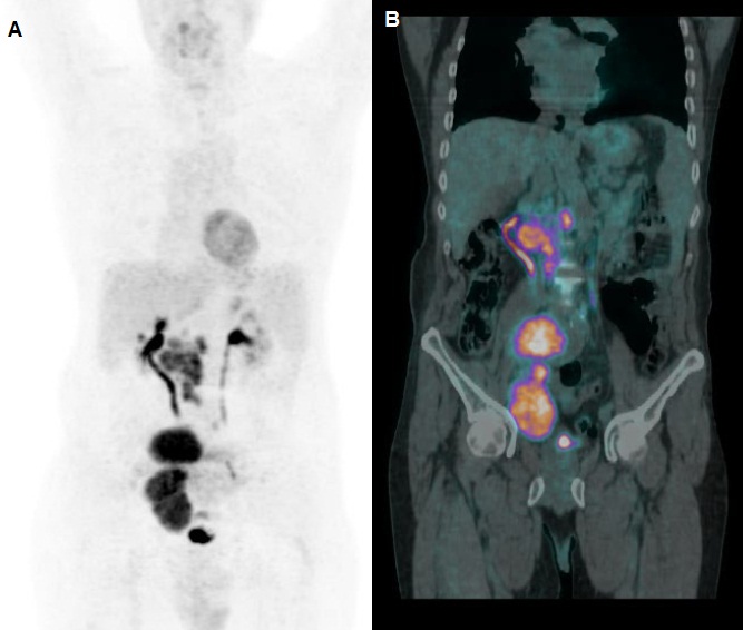
Figure 1: 18F-FDG maximum intensity projections (MIP) demonstrated intensely increased 18F-FDG uptake in large paracaval and right common iliac lymphadenopathies (A), as well shown also by the fused corresponding PET/CT coronal slices (B).
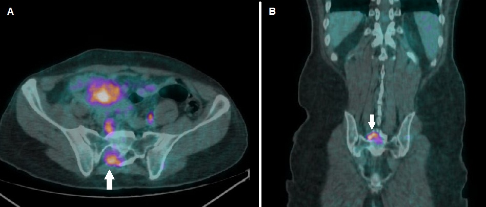
Figure 2: Fused PET/CT axial slices demonstrated focal tracer uptake located in the right sacral plexus (A, arrow), also evident in the coronal fused PET/CT images (B, arrow)
Description:
A 56 year-old male, diagnosed with diffuse large-B-cell lymphoma (DLBCL), presenting intense lumbosacral pain with irradiation to right inferior limb, was submitted to PET/CT scan with 18F-FDG as a staging study.
On the PET/CT scan an intensely increased uptake of 18F-FDG was found in correspondence of the right sacral plexus, consistent with patient’s algic symptomatology. A subsequently performed MRI confirmed the diagnosis of neurolymphomatosis. Patient was submitted to chemotherapy.
Discussion:
Neurolymphomatosis (NL) is a rare and severe clinical condition, consisting in a nervous root dysfunction secondary to infiltration by lymphoma. NL is an important prognostic factor, since the response rate to chemotherapy is of about 80% with a variable long-term outcome.
NL diagnosis, especially through conventional contrast-enhanced CT, may be challenging. In this field, the role of PET/CT with 18F-FDG, although it is still to be defined with well-designed clinical studies, seems to be promising.
Reference
Gan HK, Azad A, Cher L, Mitchell PL. Neurolymphomatosis: diagnosis, management, and outcomes in patients treated with rituximab. Neuro Oncol. 2010;12(2):212–215. doi:10.1093/neuonc/nop021
This Case was kindly provided by:
Luca Filippi, MD
Dep. of Nuclear Medicine, “Santa Maria Goretti” Hospital,
Via Canova, 04100, Latina
Italy
Contact