One stop shop:
Rectal cancer staging with PET/MRI
Patient’s history
A 59 years old male patient presented with rectal bleeding and recently pathologically proven rectal adenocarcinoma. PET/MRI was performed for local rectal staging, nodal and metastatic workup.
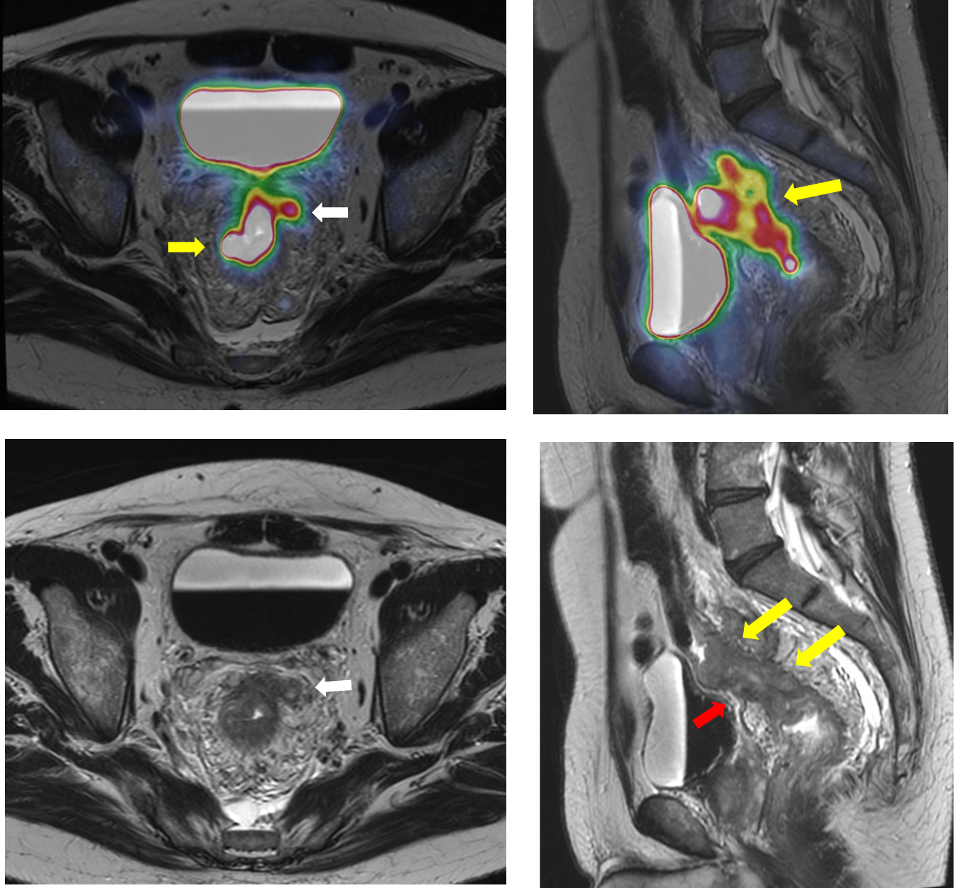
Local staging: [T]
- Circumferential irregular mural thickening is seen implicating the mid and upper rectum where this mass lesion is measuring about 6.7 x 4.5 x3.5 cm. (Yellow arrows)
- It is infiltrating the muscularis propria with multiple sizable extra mural extension into the surrounding meso-rectum, reaching a thickness of about 23mm. (White arrows)
- Significant extra mural vascular invasion is seen.
- There is infiltration of the peritoneal reflection noted at the pelvis. (red arrows)
- The mass showed:
- Significant restricted diffusion with mean ADC value of about 0.5×10⁻³ mm2 /sec.
- Increased metabolic activity reaching 30 SUVmax.
- Significant hyper-enhancement with pronounced increased K-trans value of 0.616, for the mass compared to 0.08, for the normal lower rectum.
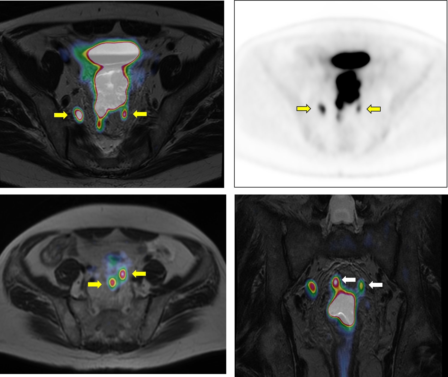
Nodal staging: [N]
- Multiple enlarged hypermetabolic lymph nodes are noted at perirectal, presacral and right internal iliac regions. (Yellow arrows)
- The perirectal lymph nodes are seen inseparable from the meso-rectal fascia on both sides. (positive circumferential resection margin) (White arrows).
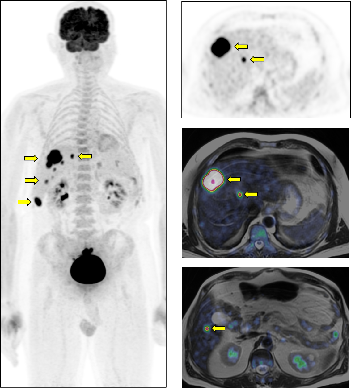
Distant Metastasis: [M]
- The liver shows multiple hypermetabolic and diffusion restricted bilobar focal lesions. (yellow arrows)
Overall staging:
- T4a, N2, M1.
This Case was kindly provided by:
 Yasser Abd El-Azim Abbas, M.D., Ph.D.
Misr Radiology Center
Department of Radiology
HCC, Off 90 St., by MRC Square,
New Cairo
Egypt
You can find more information about the Misr Radiology Center on our storyboard here.