Neuroendocrine tumor heterogeneity on hybrid imaging
INTRODUCTION:
The 68Ga-DOTATOC PET/CT is an imaging technique for detecting and characterizing neuroendocrine tumors (NETs). It allows whole-body imaging of cell surface expression of somatostatin receptors (SSTRs) and is rapidly evolving as the new imaging standard of reference for the detection and characterization of NETs. The use of 68Ga-DOTATOC PET/CT together with 18F-FDG PET/CT is complementary and help identify both well- and poorly differentiated phenotypes (tumor heterogeneity), thereby allowing tumor characterization, prognostication, and better selection of appropriate therapy for individual patients.
CASE #1
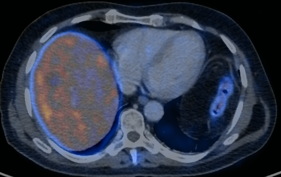
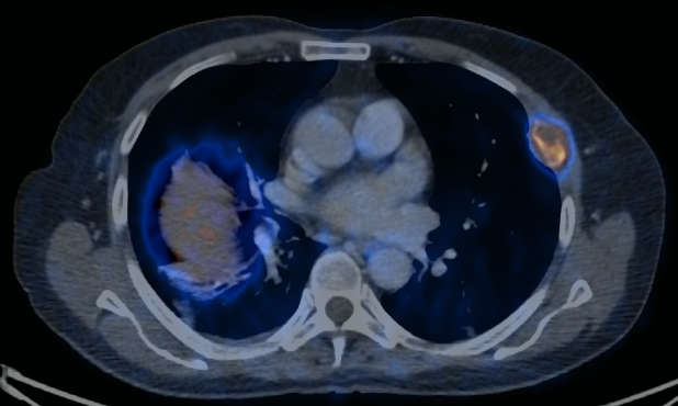
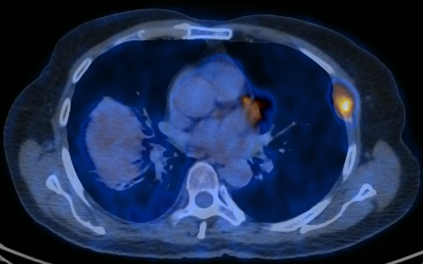
A 71 years old patient diagnosed with metastatic lung carcinoid tumor (hepatic, splenic and osseous metastasis) presents a subcarinal lymphadenopathy with different uptake patterns depending on the tracer used during the study. We have performed a comparative 18F-FDG and 68Ga-DOTATOC PET/CT study where we have noticed an increased FDG uptake in the posterior right corner of the lesion while the uptake of DOTATOC was mostly in the anterior left corner of the lesion. This probably means that the posterior right corner contains cells with high glycolytic metabolism but, at the same time, decreased SSTR expression while the anterior left corner presents low glycolytic metabolism with high levels of SSTR expression. To sum up, this adenopathy is presenting 2 different types of neuroendocrine cell populations: well differentiated in the anterior left corner and poorly differentiated in the posterior right corner.
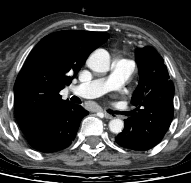
CT with contrast enhancement
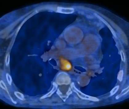
68Ga-DOTATOC PET/CT
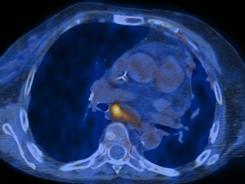
18F-FDG PET/CT
CASE #2
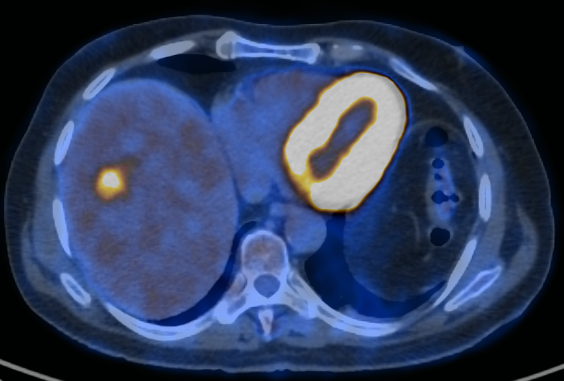
A 67 years old patient diagnosed with metastatic lung carcinoid tumor (hepatic and osseous metastasis). On the 68Ga-DOTATOC PET/CT there is no significant tracer uptake whereas on the 18F-FDG PET/CT there is one lesion in the liver segment VII that presents an increased FDG uptake. This liver lesion contains cells with high glycolytic metabolism but, at the same time, decreased SSTR expression. Unfortunately, we do not have a biopsy of this liver lesion. Anyway, we have a biopsy of one of the bone metastasis (anterior at left 5th rib), that was showing avidity for both 18F-FDG and 68Ga-DOTATOC, with a result of 20% Ki67 expression (classified as a G2 tumour). This demonstrates the heterogeneity of different NET lesions within one individual.
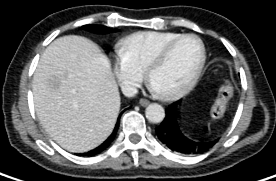
CT with contrast enhancement
68Ga-DOTATOC PET/CT
18F-FDG PET/CT
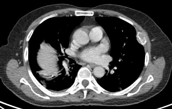
CT with contrast enhancement
68Ga-DOTATOC PET/CT
18F-FDG PET/CT
TEACHING POINT:
Tumor grade is traditionally based on the results of a single biopsy performed at the site that is most easily and safely accessible by means of percutaneous core biopsy or surgical excision. Molecular imaging, however, can allow whole-body tumor characterization, with sites of well- and poorly differentiated disease demonstrated at DOTATOC PET/CT and FDG PET/CT, respectively. The fact that different grades of disease are seen at different sites reflects tumor heterogeneity and highlights the limitations of a single random biopsy. Knowledge of this phenomenon is pivotal in guiding patient management.
This Case was kindly provided by:
Michal Pudis
Hospital Universitari de Bellvitge
Servicio de Medicina Nuclear-PET
Av. Feixa Llarga s/n
08907 L’Hospitalet de Llobregat
Barcelona, Spain
Contact