by ESHI | Feb 15, 2023 | All News, Cases
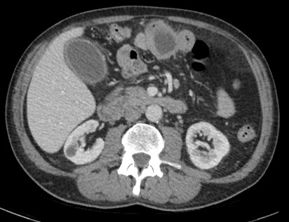
Primary intestinal diffuse large B-cell lymphoma (DLBCL) Fig. 1a Fig. 1b Fig. 1: Contrast-enhanced CT (a-c) performed in a 63-year-old male patient that presented with vomiting and fever and referring diffuse abdominal pain and unexplained weight loss in the months...
by ESHI | Apr 14, 2022 | All News, Cases
Primary carcinoid tumor of the lung Fig. 1a Fig. 1b Fig. 1c Fig. 1d Fig. 1: Unenhanced CT examinations performed by a non-smoker 71-year-old woman during follow-up for a middle lobe pulmonary nodule. The nodule was incidentally discovered during a CTU performed for...
by ESHI | May 4, 2021 | All News, Cases
Takayasu arteritis: 18F-FDG-PET/CT Figure 1. CECT examination performed in a 37 year-old woman, admitted for suspected ascending aorta ectasia suitable for surgery. Axial (a, c, d) and coronal (b) CECT images show homogeneous low density thickening of aortic wall,...
by ESHI | Mar 26, 2021 | All News, Cases
Neuroendocrine tumor heterogeneity on hybrid imaging INTRODUCTION: The 68Ga-DOTATOC PET/CT is an imaging technique for detecting and characterizing neuroendocrine tumors (NETs). It allows whole-body imaging of cell surface expression of somatostatin receptors (SSTRs)...
by ESHI | Dec 15, 2020 | All News, Cases
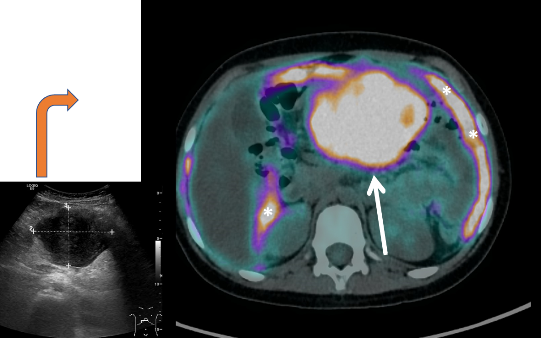
Burkitt lymphoma – 18F-FDG-PET/CT Ultrasound (US) Figure 1. Ultrasound images performed at the Emergency Room in an 8 years-old male, presenting for abdominal pain, worsening since 1 month and causing awakening over night. Fig. 1a.: Enlarged abdominal lymph...
by ESHI | Oct 23, 2020 | All News, Cases
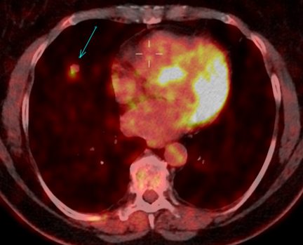
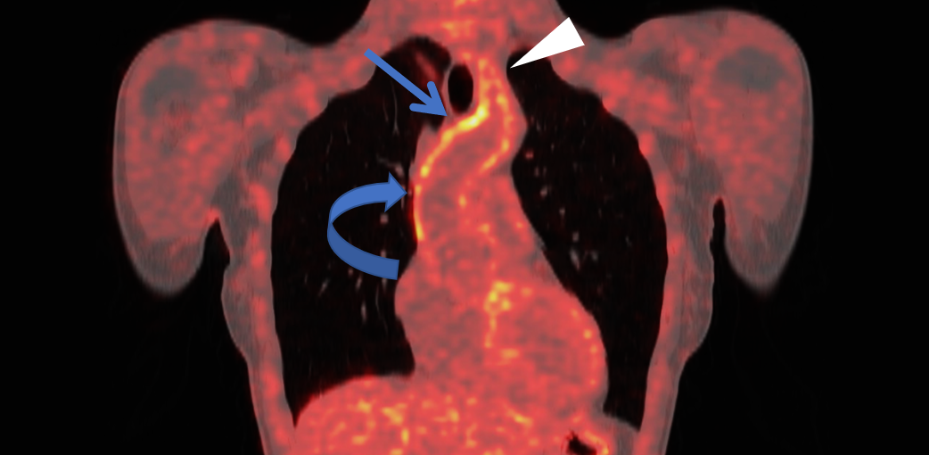
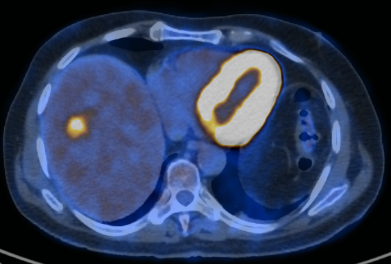
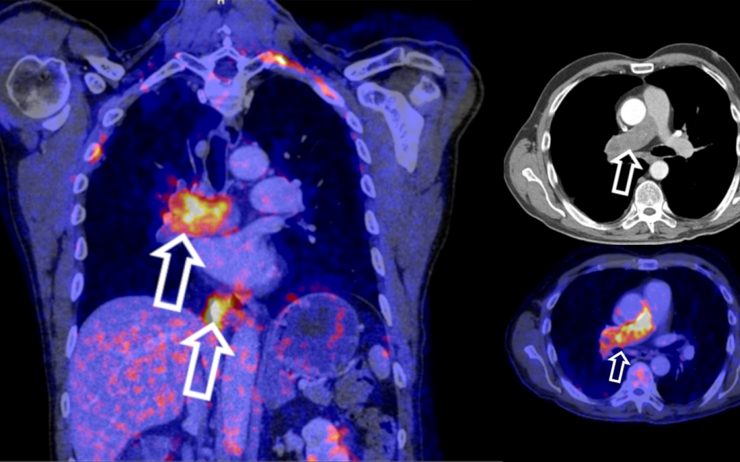
Intimal sarcoma of the right pulmonary artery: 18F-FDG PET/CT A 54-y/o male patient presented with dyspnoea and right-sided thoracic pain radiating to the right arm. After exclusion of myocardial infarction, computed tomography revealed a contrast filling defect of...